Clinical and laboratorial characteristics associated with rickettsiosis
Karina Citlalli Sandoval-Duarte† and Porfirio Felipe Hernández-Bautista*
*Coordinación de Calidad de Insumos y Laboratorios Especializados, Instituto Mexicano del Seguro Social, Ciudad de México 07760, México.
Abstract
Introduction: Rickettsiosis is transmitted by arthropod vectors (ticks, mites, lice and fleas), caused by bacteria of the genus Rickettsia of the Rickettsiaceae family, and represents a serious impact on worldwide health. The disease can affect anyone, two-thirds of cases occur in children under 15, the highest incidence being in those between 5 and 9, who have increased risk of complications, and currently up to 3% of them in the U.S. die.Objetive: To determine whether epidemiological, clinical and laboratory characteristics exist associated with the diagnosis of rickettsiosis in the Hospital General Regional (HGR) No. 1 de Cd. Obregón, Sonora, Mexico.
Materials and methods: Epidemiological study of cases and controls. Cases included were those patients with clinical symptoms with positive results in any serological tests; as controls patients with clinical symptoms who had negative results in any serological tests, presented between the 1st of January 2008 and the 31st of December 2013. Analysis was carried out for quantitative variables obtained using parametric measurements or non-parametric depending on their distribution; proportions for qualitative variables. It be calculated using the Odds Ratio.
Results: This case-control study in Sonora identified rash as a key risk factor for rickettsiosis (OR = 2.71, 95% CI 1.3–5.65). In complete blood counts, the main significant risk factors were low hematocrit (OR = 5.45, 95% CI 1.03–28.76), low hemoglobin (OR = 2.15, 95% CI 1.05–4.36), and neutrophilia (OR = 2.62, 95% CI 1.10–6.94). Having received a previous misdiagnosis (OR = 3.7, 95% CI 1.06–12.81) also increased the risk of complications and hospitalization. Finally, monocytosis acted as the only significant protective factor (OR = 0.28, 95%IC 0.08-0.78) against the disease.
Conclusions: This study highlights the need for ongoing education of medical personnel in Sonora. Rickettsiosis should not be diagnosed solely based on rash or bites, as this delays treatment. The typical advanced profile includes rash, low hematocrit, and a previous misdiagnosis. Despite limitations in older records, the multivariate model is robust.
Keywords: Rickettsiosis, clinical symptoms, hematic biometry, diagnosis, treatment.
INTRODUCTION
Rickettsiosis, transmitted by arthropod vectors mainly ticks, mites, lice and fleas, is a disease caused by bacteria of the genus Rickettsia which belongs to the Rickettsiaceae family, and represents a serious impact on worldwide health. These pathogens are responsible for numerous outbreaks worldwide and are endemic diseases in America, parts of Europe and Africa.1,2,3,4
Rickettsia are obligate intracellular bacteria they infect the endothelial cells of blood vessels. The most commonly used classification divides them into the spotted fever group, which contains the species R. rickettsii, R. conorii and R. peacockii among others, the Typhus Group (TG) in which R. typhi and R. prowazekii are found, the transitional group (TRG) composed of R. felis and R. akari and finally, the ancestral group (AG) which includes R. canadensis and R. bellii.2,3,5
In Mexico, as reported by the ‘Sistema Nacional de Vigilancia Epidemiologica’ (SiNaVE) (National System of Epidemiological Observation), the epidemic typhus caused by R. prowazekii that occurs principally in individuals living in overcrowded and unsanitary conditions, is chiefly Rickettsiosis. Following this, murine typhus Rickettsia is the second largest in the country. The third largest of Rickettsiosis corresponds to spotted fever caused by R. rickettsii and has the highest national mortality rate.6
The disease can affect anyone, two-thirds of cases occur in children under 15, the highest incidence being in those between 5 and 9, who have increased risk of complications, and currently up to 3% of them in the U.S. die.7
Patients present clinical symptoms characterized by fever, headache, signs such as rash, muscle pain, digestive symptoms (abdominal pain, nausea, vomiting), neurological signs, hemorrhagic manifestations impaired Hematic biometry (thrombocytopenia, leukocytosis, neutrophilia, elevated transaminases), and a history of tick bites, lice or fleas.4,5,8,9,10
Furthermore, there are factors that contribute to endemicity and interfere with the optimal implementation of control measures such as: poverty, ignorance, dirt, poor housing, poor access to communities, limited resources: economic, trained personnel, transportation, etc.4,5,10,11
These diseases are gaining recognition among physicians as an important set and often indistinguishable due to diagnostic problems and clinical management, such as late diagnosis or lack of diagnosis, which can generate prolonged hospital stays, intensive care and in extreme cases, deaths in up to 53% of cases. If treatment with Tetracycline or Chloramphenicol started early (within the first 5 days of illness) mortality was significantly reduced to 20 or 5%.1,4,10,11
MATERIALS AND METHODS
A study of cases and controls was carried out in HGR No.1 in Obregon, Sonora. The study population was taken from hospital records and outpatients between 1st January 2008 and 31st December 2012. They are defined as everyone with clinical symptoms and positive laboratory results for Rickettsiosis or by epidemiological association with another confirmed case. The control was those with suspected Rickettsiosis who has been excluded in laboratory or epidemiological association could not be proven to have a confirmed case. Regarding the ethical side of the investigation, according to the Regulations of General Health Law in Health Research, this investigation is judged to be of minimal risk.
Inclusion criteria were case patients with clinical symptoms and positive results for Rickettsiosis through detection by Indirect immunofluorescence of IgM and IgG antibodies or it was proved by epidemiological association with an already confirmed case; and as a control anyone with suspected Rickettsiosis who has been proven to not be infected as ruled by the same laboratory techniques, but were positive for other infectious or immunological diseases (eg Dengue, Leptospirosis, Brucellosis, Leukemia, etc.). Elimination criteria for both cases were that epidemiological studies could not be located or did not comply with 80% of the information.
Proceeding the list of assumed patients being obtained, application and verification of the Nominal of the Ministry of Health, the results of diagnostic tests for Rickettsiosis were made by the State Laboratory of Public Health (LESP), ruling and association with other confirmed cases; and the results subrogated by the Mexican American Hospital Laboratory Diagnostics and were allocated to the Epidemiology Service. This meant patients could be classified according to the result. Confirmatory tests consisted of the detection of IgM and IgG antibodies by micro-agglutination, complement fixation, indirect immunofluorescence by isolation of Rickettsia from blood or observation of smears.
Once classified, patients proceeded to obtain epidemiological studies of Vector-Borne (ETV) performed by Disease Epidemiology Service, as well as medical records were reviewed to supplement information and results of the first biometrics blood count and liver function tests at the time of suspicion of infection were obtained, patient outcome and treatment received.
With the information obtained a univariate analysis was conducted, wherein the means of quantitative variables and proportions for qualitative variables were attained. In bivariate analysis for qualitative variables Chi Square, Fisher’s Exact Test or Z Test as appropriate and for quantitative variables Student T-Test or Mann-Whitney were used. An Odds Ratio was calculated as a measure of risk considering statistically significant variables those that yielded P <0.05 with confidence intervals at 95%. The multivariate analysis was done using non-conditional logistical regression. The backward Euler method was used for selecting variables in the regression according to their explanatory capacity. The analysis was conducted with STATA 12 statistical software.
RESULTS
During the study period 374 patients with clinical symptoms of rickettsiosis, in accordance with hospital records and external consultation, were identified. Of these, 244 corresponding epidemiological studies of vector-borne disease and 140 dossiers were found; a total of 44 patients were eliminated for not meeting the selection criteria, finally leaving a case-control ratio of 1:3 (50 cases and 150 controls).
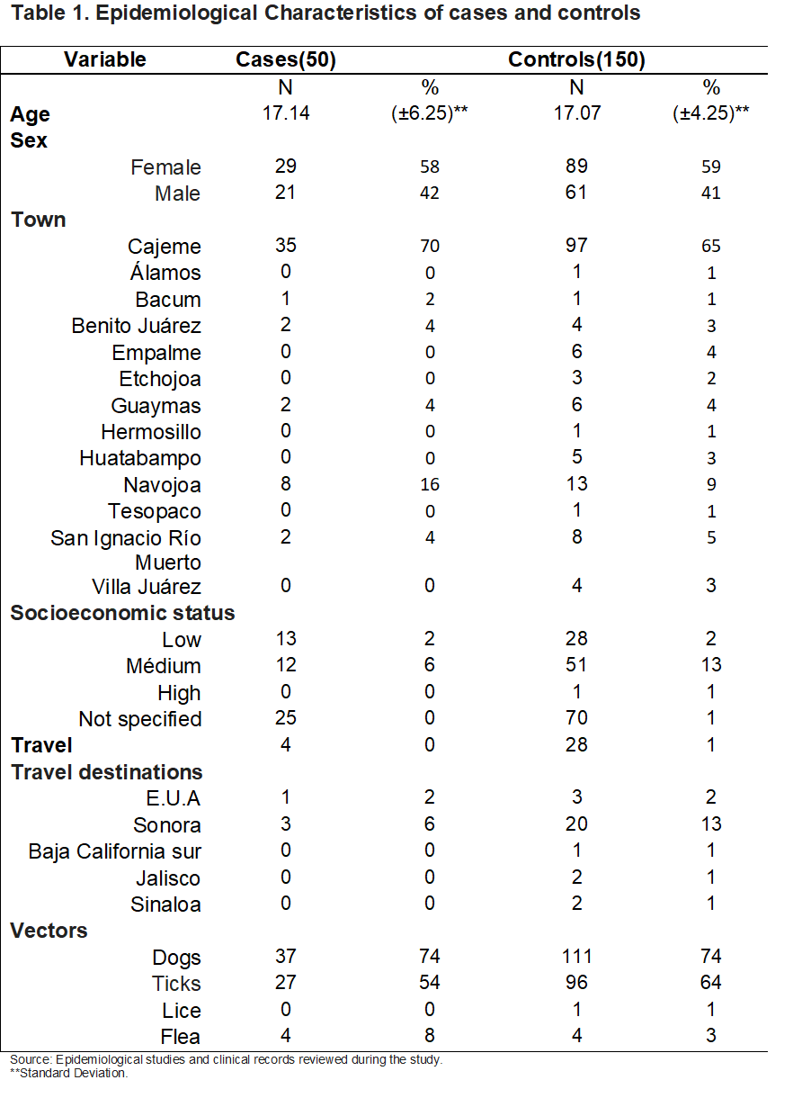
According to the epidemiological characteristics, the mean age was no different between the groups, with respect to gender greater frequency was witnessed in females in both groups. The three most affected municipalities presenting the greatest number of controls are Cajeme, Navojoa and San Ignacio Río Muerto. A total of 32 patients had traveled at least one month before the onset of symptoms; the most frequently visited locations were within the state of Sonora and were primarily visited by patients in the control group. The same percentage of the control group members lived with dogs. Among the vectors, ticks were the most prevalent in both groups, followed by fleas. (Table 1).
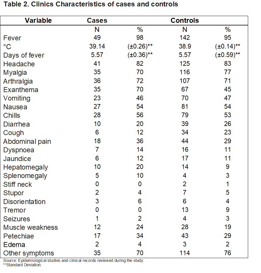
In terms of clinical symptoms, a large proportion of patients experienced a fever of 39°Con average. The duration of persistence of fever was equal in both groups, 5.5 days. As described in the literature, fever, headache, myalgia and arthralgia form the principal symptoms leading to the suspicion of Rickettsiosis, coupled with the presence of vectors, which could be demonstrated in this study. In as far as Petechia occurred in 34% of cases and 29% of the controls, this expiring will not investigate the characteristics of palms and soles.
Only one case presented seizures associated with a fever. Among other symptoms described by patients, headache (82%), arthralgia (72%), exanthema (70%), chills (56%) and nausea (27%) were most frequent in both groups. Bleeding data at any level, positive tourniquet tests and mottled skin usually associated with dengue hemorrhagic fever occurred mainly in the controls (Table 2).
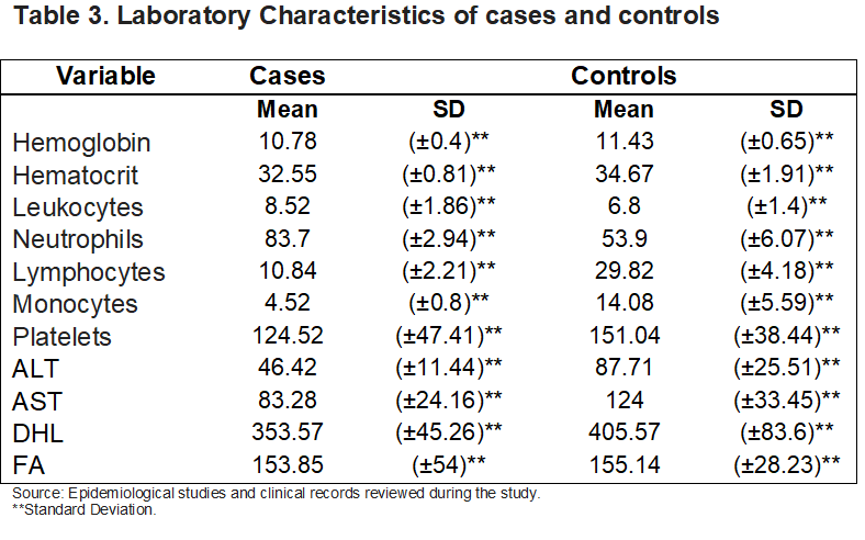
The meaning of the hematic biometry and liver function tests are presented in Table 3. As can be seen in both cases all patients had hypochromic anemia with leukocyte within normal ranges. The presence of differential neutrophils in the cases and monocytosis in the controls were also observed. With regards to the platelets, it is evident that the cases present with thrombocytopenia, while the controls showed an average within normal ranges. The liver function tests are elevated in both groups, however in the case of controls, the increase is more significant.
As follow up and patient outcomes (Table 4), 92 of them received first level health care after the suspicion of Rickettsiosis; of these 92, 62 were primarily diagnosed with Pharyngitis, urinary tract infection, vector-borne disease (VTE) mainly Dengue or some other disease of the 30 patients remaining the diagnosis is unknown because it was not specified in the medical notes or the medical records couldn’t be found.
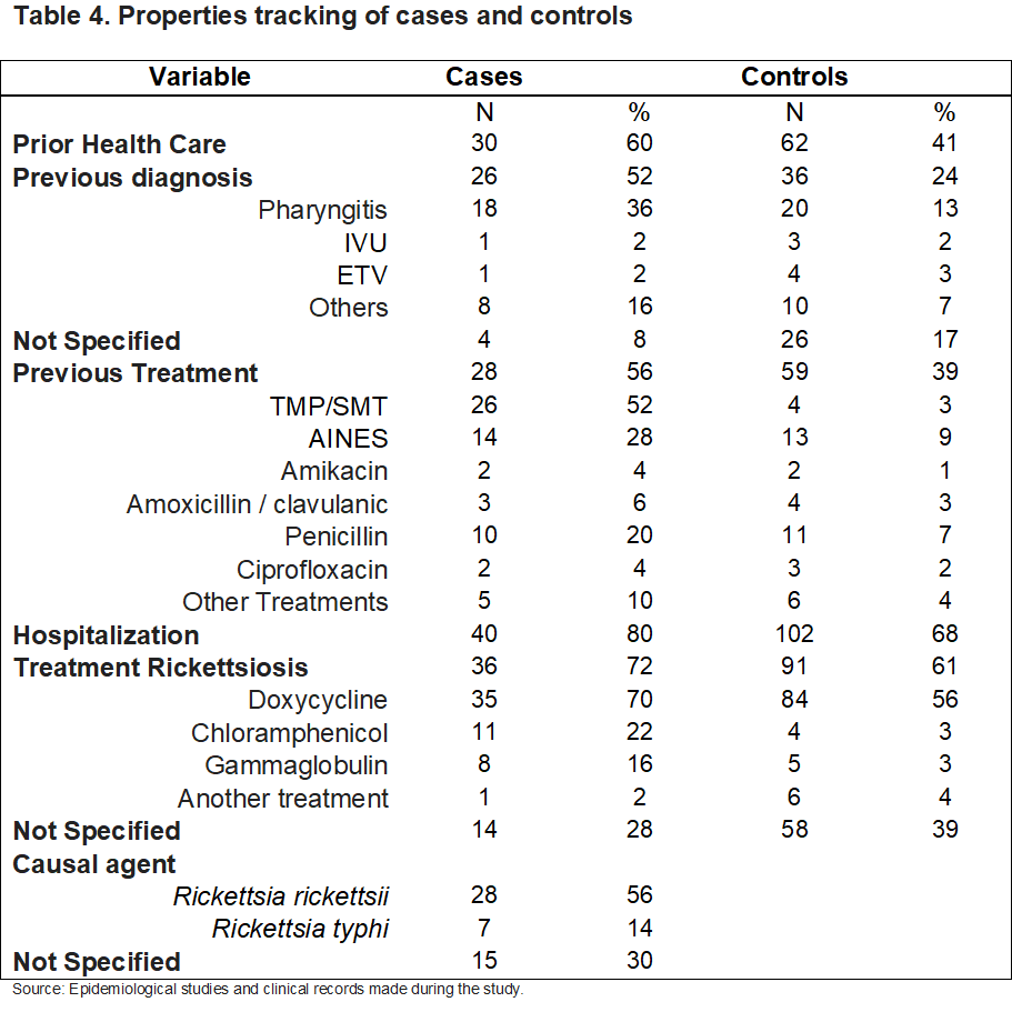
Of the 92 patients, only 87 received some form of treatment, mainly with antibiotics such as trimethoprim-sulfamethoxazole and some NSAIDs. Once patients came to HGR, and there was a suspicion of the symptoms of Rickettsiosis, they were assessed by the Epidemiology Service, who initiated protocol for this disease, conducting an epidemiological study for VTE, also taking a serum sample to determine the diagnosis; 142 patients required hospitalization (40 cases and 102 controls).
In 72 of patients the type of treatment received is unknown, as no information was found. Only one of the controls received no treatment for this infection, because although it was suspected rickettsioses, the clinical doctor began medical treatment for another illness, which was verified by clinical analysis. Of the cases the causal agents diagnosed were Rickettsia rickettsii, Rickettsia typhi, it is noteworthy that of these, 3 cases were diagnosed with 2 causative agents (2 cases of Rickettsia rickettsii and Rickettsia typhi). In 13 cases the causative agent is not specified in nominal results of the State Laboratory of Public Health, these correspond to the first study year.
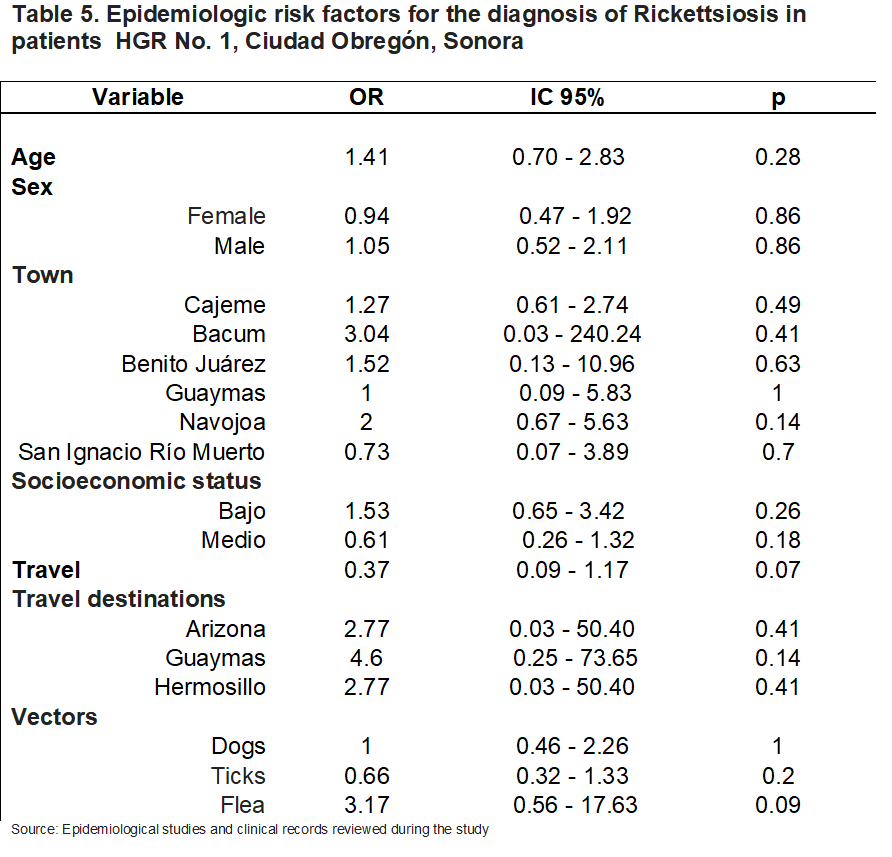
The Odds Ratios (OR) was used for the analysis of risk factors associated with the diagnosis of Rickettsiosis; in the case of epidemiological factors (Table 5) according to the confidence intervals (CI) at 95% and the value of p, none of the studied variables were statistically significant, being undetermined factors. As for the clinical picture (Table 6), it was found that exanthem is a risk factor (OR = 2.89 [95% CI 1.39- 6.17]) with a significant p of 0.0019; the rest of the symptoms are undetermined.
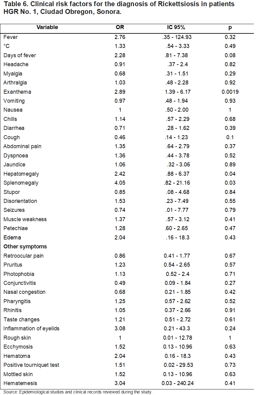
The findings in the hematic biometry (Table 7) those that are at risk are low hemoglobins (OR = 2.15 [95% CI % 1.05-4.36]), low hematocrits (OR = 2.78 [95% CI 1.37-5.67]) with significant p 0.02 and 0.0018 respectively; as well as the presence of neutrophilia with an OR of 2.62 (95% CI [1.10 - 6.94]). A lymphocyte count within normal ranges also forms a risk factor (OR = 2.39 [95% CI % 1.16-5.05]) and the presence of monocytosis is a protective factor for Rickettsiosis (OR = 0.28) with CI 95% 0.08 - 0.78. Although in liver function tests ALT, AST and LDH have 1 or higher than 1, according to confidence intervals and p are undetermined factors.
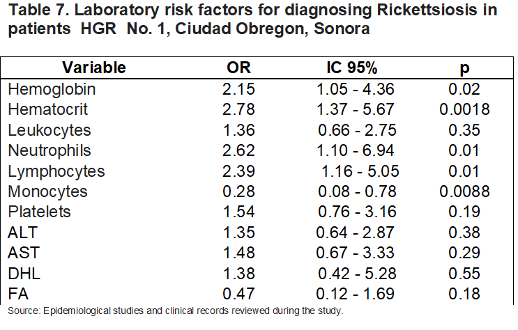
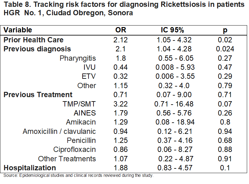
Associated factors were also evaluated to monitor patients (Table 8), verifying that many have received medical care (OR = 2.12 [95% CI 1.05-4.32]) and a previous diagnosis (OR = 2.1 [95% CI 1.04-4.28]) for the suspicion Rickettsiosis are risk factors, thereby not having received prior treatment; when the analysis is done separately from the diagnosis and no treatment it is significant that hospitalization is required.
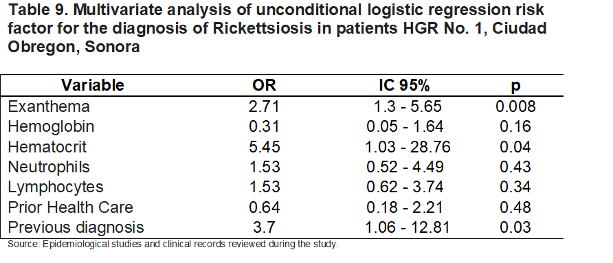
In the model of unconditional logistic regression (Table 9) exanthemata’s identified as a factor of risk with OR = 2.71 (95% CI [1.3 - 5.65]), low hematocrit OR = 5.45 (95% CI [1.03 - 28.76]) and having received a previous diagnosis OR = 3.7 (95% CI [1.06 - 12.81]); the rest of the factors are indeterminate.
DISCUSSION
This study evaluates the clinical, epidemiological, and laboratory factors associated with rickettsiosis at Regional General Hospital No. 1 in Ciudad Obregón, Sonora, a region historically vulnerable to vector-borne diseases. Our statistical findings confirm that delays in accurate diagnosis, the presence of rash, and specific hematological abnormalities are critical determinants in the disease's progression and management.
Nationally, the National Epidemiological Surveillance System (SiNaVE) indicates that spotted fever caused by *R. rickettsii* has the highest mortality rate in Mexico.6 Although the literature strongly associates disease persistence with poverty, overcrowding, and substandard housing,4,5,10 in our univariate analysis and odds ratio (OR) analysis, environmental variables did not reach statistical significance (P > 0.05). This can be explained by the high level of information bias and missing data in outpatient records, which limited accurate socioeconomic stratification.
However, it is noteworthy that 55.5% of the control group lived with dogs, compared to only 18.5% of the case group. This seemingly paradoxical finding often reflects a bias in medical suspicion: healthcare personnel tend to suspect and protocolize rickettsiosis more frequently in patients who report direct contact with dogs or ticks, sending them to the screening group (which ultimately tests negative or becomes a control group), while true cases sometimes lack this clear history in their initial interview.
Fever (average of 39°C) and headaches were consistently present in both groups, confirming the clinical challenge of differentiating this disease from other endemic conditions such as dengue.4,8 However, multivariate analysis using unconditional logistic regression identified the rash as a robust and statistically significant risk factor (OR = 2.71; 95% CI: 1.3–5.65; P = 0.0019). This is consistent with the findings of Bukowski and Paddock,8 who identified the rash as the cardinal sign of infectious vasculitis due to endothelial damage. Unfortunately, the late appearance of this sign often delays initial clinical suspicion.5,9
In the laboratory profile, the logistic regression model showed that a low hematocrit represents a critical risk factor (OR = 5.45; 95% CI: 1.03–28.76). Hypochromic anemia and decreased hematocrit reflect the severity of the microangiopathy and the possible occult hemorrhagic manifestations caused by bacterial replication in endothelial cells.3,9
Likewise, the presence of neutrophilia (OR = 2.62) was established as a strong predictor of active rickettsial infection, in contrast to the control group where monocytosis acted as a protective factor (OR = 0.28), pointing the diagnosis toward non-rickettsial viral or immunological etiologies.8 Although thrombocytopenia was evident in the average of cases, in the individual risk analysis it behaved indeterminately due to the overlap with control patients suffering from dengue, an entity that also presents with severe platelet destruction.
The most alarming findings of our research lies in the impact of the patient's journey through the healthcare system. Having received prior medical attention (OR = 2.12) and an initial misdiagnosis (OR = 2.1 in the bivariate model; OR = 3.7 in the multivariate model) were identified as significant risk factors for disease progression.
Fifty percent of the patients who sought care at a primary care level were misdiagnosed with pharyngitis and treated with ineffective regimens (such as trimethoprim-sulfamethoxazole). This aligns with the warnings of Sahni et al.4 and Astudillo-Hernández,10 who link the lack of training of primary care personnel with prolonged hospitalizations and fatal outcomes. International literature demonstrates that initiating tetracyclines (such as doxycycline) within the first 5 days drastically reduces mortality from 53% to less than 5%.1,11 In our study, diagnostic error at first contact delayed specific therapy, leading to a high hospitalization rate (40 cases) due to systemic progression of the infection before admission to General Regional Hospital No. 1.
This study highlights the critical need to implement continuing medical education programs in Sonora. Rickettsiosis should not be diagnosed solely based on the presence of a rash or a history of vector bite, as waiting for these signs perpetuates treatment delays. Limitations of the study include data loss in older clinical records and the difficulty in identifying the specific etiological agent in the early years of the sample. However, the robustness of the multivariate model demonstrates that a patient with a rash, decreased hematocrit, and a previous misdiagnosis represents the typical profile of advanced rickettsiosis requiring immediate medical intervention.
REFERENCES
1. Parola, P., Paddock, C. D., Socolovschi, C., Labruna, M. B., Mediannikov, O., Kernif, T., ... & Raoult, D. (2013). Update on tick-borne rickettsioses worldwide: a geographical approach. Clinical Microbiology Reviews, 26(4), 657-702.
2. Weinert, L. A., Werren, J. H., Aebi, A., Stone, G. N., & Jiggins, F. M. (2009). Evolution and diversity of Rickettsia bacteria. BMC Biology, 7(1), 6.
3. Gillespie, J. J., Williams, K., Shukla, M., Snyder, E. E., Nordberg, E. K., Ceraul, S. M., ... & Sobral, B. W. (2008). Genomica de Rickettsia: el impacto de la reducción genómica en la evolución de las bacterias intracelulares obligadas. PLOS ONE, 3(3), e1718.
4. Sahni, S. K., Narra, H. P., Sailesh, K., & Walker, D. H. (2019). Rickettsial diseases: progress and challenges. Future Microbiology, 14(14), 1183-1186.
5. Blanton, L. S. (2019). The rickettsioses: a practical review for clinicians. The American Journal of Medicine, 132(7), 792-799.
6. Secretaría de Salud / Sistema Nacional de Vigilancia Epidemiológica (SiNaVE). (2023). Manual de Procedimientos Estandarizados para la Vigilancia Epidemiológica de las Enfermedades Transmitidas por Vectores. Ciudad de México, México: Dirección General de Epidemiología.
7. Biggs, H. M., Behravesh, C. B., Bradley, K. K., Dahlgren, F. S., Drexler, N. A., Dumler, J. S., ... & Traeger, M. S. (2016). Diagnosis and management of tickborne rickettsial diseases: Rocky Mountain spotted fever and other spotted fevers—United States: a practical guide for health care professionals. MMWR Recommendations and Reports, 65(2), 1-22.
8. Bukowski, D. M., & Paddock, C. D. (2020). Clinical and laboratory manifestations of spotted fever group rickettsioses. Journal of Intensive Care Medicine, 35(11), 1105-1115.
9. Álvarez-Hernández, G., Murillo-Benítez, C., Candia-Plata, M. D. C., & Moro, M. (2015). Clinical features and predictors of fatality in New World spotted fever. Biomédica, 35(1), 98-105.
10. Astudillo-Hernández, M., Flora-Flores, M. G., Vences-Blanco, M. A., & Olivera-Díaz, H. (2018). Factores socioeconómicos, pobreza y rickettsiosis: desafíos en salud pública en regiones endémicas. Revista Panamericana de Salud Pública, 42, e104.
11. Todd, S. R., Dahlgren, F. S., Traeger, M. S., Beltrán-Aguilar, E. D., Paddock, C. D., Regnery, R. L., & Childs, J. E. (2015). No evidence of tooth staining in children treated with doxycycline for suspected Rocky Mountain spotted fever. The Journal of Pediatrics, 166(5), 1246-1251.
Citation
Sandoval-Duarte KC† and Hernández-Bautista PF, Clinical and laboratorial characteristics associated with rickettsiosis, ERSJ 2026,1(5) 41-54
Academic Editor: Ana Karem Ramírez Utrera , Received: June-11-2026, Revised: June-22-2026, Accepted: July-06-2026, Published: July-18-2026.