Epidemiological characteristics in patients diagnosed with Rocky Mountain Spotted Fever
Susana Patricia Cantú-González and Eduardo Arturo Serrano-García
Hospital General de Zona No. 14, Instituto Mexicano del Seguro Social, C. República de Cuba, El Mirasoles, 83120 Hermosillo, Sonora.
Abstract
Introduction: Rocky Mountain Spotted Fever (RMSF) is an endemic infectious disease with a nonspecific clinical presentation and a high mortality rate in the state of Sonora. This research aims to describe the main epidemiological, clinical, and laboratory aspects of the disease in order to provide timely diagnosis and treatment to patients served by General Hospital Zone 14 in Sonora.Objetive: To describe the epidemiological, clinical and laboratory characteristics in patients diagnosed with Rocky Mountain Spotted Fever treated at General Hospital Zone 14, Sonora during the period from January 2018 to March 2023.
Materials and methods: A descriptive, cross-sectional study was conducted on patients admitted to General Hospital Zone 14 based on epidemiological studies and a nominal census of vector-borne disease and electronic records.
Results: Fifty-nine percent of cases were in females, with the highest proportion between 18 and 64 years of age. Contact with the vector, prior medical attention, and initiation of doxycycline ≥ 6 days prior to symptom onset were the predominant risk factors in this study. The classic triad was present in only 38% of cases, and paraclinical abnormalities were observed within the first 72 hours of symptom onset.
Conclusions: The epidemiological findings were consistent with other current studies. The symptoms presented were nonspecific, and a case fatality rate of 43% was obtained. It is important to recognize the diagnosis even when exposure to the vector is unknown, considering the high prevalence and lethality of the disease in the state of Sonora.
Keywords: Fever, spotted, rickettsia rickettsi, rickettsiosis.
INTRODUCTION
Rocky Mountain Spotted Fever (RMSF) is a re-emerging infectious disease with a 50% case fatality rate in the state of Sonora. Currently, the clinical presentation is known to be nonspecific in the first few days of onset, so the diagnosis is not always suspected in the acute stage of the disease. Consequently, the treatment of choice is administered late, when the disease is already advanced and carries a higher risk of a fatal outcome, as has been observed in recent studies.
RMSF is an infectious disease caused by the bacterial species Rickettsia rickettsii. Historically, cases of a febrile illness with nonspecific clinical characteristics, including a petechial rash, headache, and high fever, have been reported in the states of Sonora and Sinaloa, reaching a high case fatality rate. Since then, the epidemiological, clinical, and entomological aspects of this disease in Mexico have been further detailed. 1
It is now known that the disease is transmitted through the bite of one of the most important vectors in northern Mexico, the tick Rhipicephalus sanguineos sensu lato, which is abundant on its main host, the stray dog. The rapid increase in the number of these hosts has led to a growing understanding of the epidemiological and ecological circumstances that give rise to outbreaks in regions of the southeastern United States (USA) bordering Mexico.1
The Rickettsiaceae family consists of the genera Rickettsia and Orientia. The American pathologist Ricketts first described the genus Rickettsia in 1909. This genus comprises four groups, including the species with the greatest epidemiological impact in our country: R. rickettsii (MRSM) from the spotted fever group and the typhus group, R. typhi (murine typhus), and R. prowazekii (epidemic typhus). The genus Orientia includes one species, O. tsutugamush.2
The bacteria are small coccobacilli, measuring no more than 0.3 to 0.5 microns, are obligate intracellular parasites, and grow only in the cytoplasm of eukaryotic cells. The genus Rickettsia possesses a minimal peptidoglycan layer, which is why it stains weakly, and its lipopolysaccharide (LPS) has only weak endotoxin activity. The outer membrane protein A (OmpA) is expressed on the surface of R. rickettsia and is responsible for its ability to adhere to endothelial cells, enter, escape the phagosome, and multiply in the cytoplasm and nucleus before exiting the cell to migrate to the next.3
In Mexico, the brown dog tick, Rhipicephalus sanguineus, belonging to the family Ixodidae, is the main reservoir and vector. It feeds on dogs and can persist in urban environments. In the United States, the dog tick (Dermacentor variabilis) is prevalent, while the wood tick (Dermacentor andersoni) is found in the Rocky Mountain states and southeastern Canada. They feed on various mammals, including wild rodents, and transmission is most common during activities in rural areas where prolonged exposure to the tick occurs, typically 6 hours or more.4
Approximately 1 to 3% of ticks become infected with R. rickettsii transovarially or through feeding on infected animals. Once infected, these ticks do not die and serve as reservoirs and vectors for the species. Subsequently, transmission to humans occurs through the tick's saliva during a bite when it remains attached to the skin and feeds for 12 to 24 hours (although transmission through exposure to tick fluids or feces has also been described).5
After an incubation period of 2 to 14 days, the bacteria spread via the lymphatic and circulatory systems. Most species adhere to their target cell, the endothelial cell, escaping the phagosome and proliferating intracellularly to the point of rapidly spreading from cell to cell. Other species, such as R. akari, responsible for rickettsial pox transmitted by infected mites, target macrophages. This intracellular location of Rickettsia spp. is what makes direct detection of the organism in the laboratory difficult.6
Rickettsia organisms have been found on every continent except Antarctica. Most species are prevalent in certain regions due to climatic, vector, and host conditions. They can affect people of any age; however, in the United States, two-thirds of cases occur in children under 15 years of age, with the highest incidence and greatest risk of complications in children between 5 and 9 years old. In Sonora, in 2012, the case fatality rate was reported to be between 8% and 17.8%, with an upward trend since the disease's reemergence at the beginning of the decade.7
Mexican spotted fever (MSF) represents a significant disease burden that continues to be underdiagnosed globally. Clinical and laboratory diagnosis is challenging, and a confirmatory result should not be awaited before initiating treatment when the disease is suspected. R. rickettsii is the most lethal species among spotted fever infections, and delays in diagnosis and timely treatment (within the first five days of symptom onset) with the antibiotic of choice, doxycycline, endangering the lives of the population, particularly the most affected age group, children.5
Delayed initiation of antibiotic therapy carries a fourfold increased risk of mortality compared to initiating treatment before the sixth day of symptom onset. Therefore, it is important to keep healthcare personnel updated on the characterization of symptoms, clinical course, and laboratory parameters of this disease. Likewise, to educate the community on the principles of primary and secondary prevention.5
Therefore, the objective of the study was to describe the epidemiological, clinical, and laboratory characteristics of patients diagnosed with Rocky Mountain Spotted Fever treated at General Hospital Zone 14, Sonora, during the period from January 2018 to March 2023.
MATERIALS AND METHODS
A descriptive study was conducted at General Hospital No. 14 with a Burn Unit. The study period was from January 1, 2018, to March 31, 2023. Patients of any age and sex were included. They underwent an epidemiological study of vector-borne diseases (VBDs) and were reported as probable cases of Rickettsiosis based on symptoms, clinical course, and suggestive laboratory profile upon admission. Subsequently, they were diagnosed with Rickettsia spp. and Rickettsia rickettsii according to the 2022 Guidelines for Laboratory Surveillance of Rickettsiosis, which include tests to detect Rickettsia spp. by real-time reverse transcription polymerase chain reaction (RT-PCR) and determination of Rickettsia rickettsii by the InDRE by quantitative PCR (qPCR) and indirect immunofluorescence for the detection of anti-IgG antibodies against spotted fever group rickettsiae (antigen: R. rickettsii), taking into account the cases as confirmed by this last test those with the first positive serological sample and then, at least 2 weeks later, a second serum sample with seroconversion greater than or equal to four times the titer of the first sample. Patients without a confirmatory diagnostic test were excluded. Patients with negative results for rickettsiosis were excluded, as were those with incomplete epidemiological studies.
The main variables studied were sex, age, exposure history, epidemiological history, clinical course, and timeliness of treatment. Permission was previously requested from General Hospital Zone 14 to use information from its database and electronic records exclusively for data review and collection. Information was obtained from the nominal census and the epidemiological study of vector-borne diseases (VBDs), electronic records, and the IMSS HGZ 14 laboratory platform. An Excel database was created to list the variables under study. Subsequently, the SPSS statistical software (Statistical Package for the Social Sciences) was used to analyze the corresponding variables.
A descriptive analysis was performed for the qualitative variables, obtaining proportions, measures of morbidity frequency, calculating point prevalence, and the case fatality rate. For the quantitative variables, an analysis of measures of central tendency and dispersion was performed, obtaining the mean and standard deviation, respectively.
RESULTS
For this study, 214 probable cases that received medical attention at General Hospital Zone 14 in Hermosillo, Sonora, Mexico, from January 2018 to March 2023 were considered, of which 68 met the inclusion criteria. 146 cases were excluded due to a negative PCR test result and a single positive IgG IFA test result; no cases were excluded from this study.
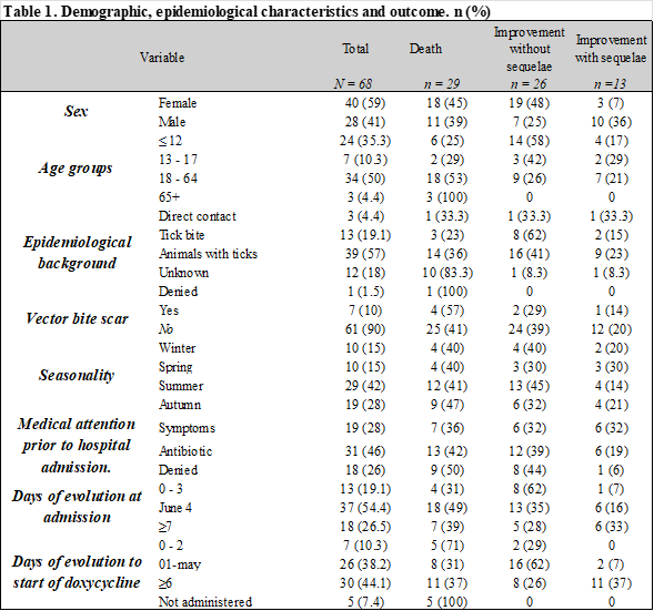
In accordance with the objectives of this study, the following results were obtained: 59% (n=40) of the cases were female and 41% (n=28) were male. The age group with the highest proportion of cases was 18 to 64 years, with 50% (n=34), followed by the ≤ 12 years group with 35% (n=24%). Fifty-seven percent (n=39) of cases involved pets in the home and/or stray dogs outside their homes carrying ticks; 19% (n=13) reported a tick bite 14 days prior to the onset of symptoms; and in 10% (n=7), a tick bite scar was found during the physical examination. The season with the highest incidence of cases was summer (June-September), accounting for 42% (n=29) of cases. Forty-six percent (n=31) of cases received medical consultation (public or private) with a prescription for antibiotic therapy other than doxycycline prior to hospital admission, and 42% (n=13) of these cases resulted in a fatal outcome. Hospital admission occurred most frequently between 4 and 6 days after the onset of symptoms, representing 54% (n=37) of cases. Likewise, in 44% of cases, doxycycline treatment was initiated ≥ 6 days after the onset of symptoms. Table 1
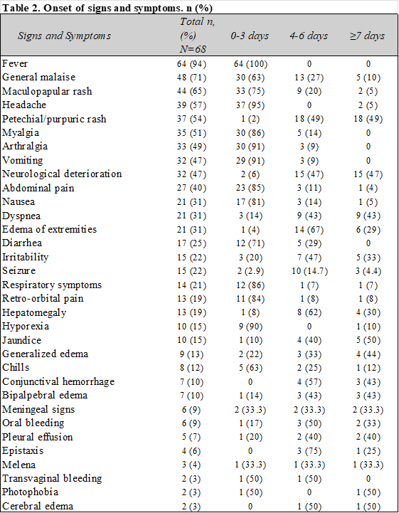
The main signs and symptoms that appeared were fever 94% (n=64), malaise 71% (n=30), maculopapular rash 65% (n=44), and headache 57% (n=39), which predominated in the first 3 days of the onset of symptoms, accompanied by gastrointestinal symptoms such as vomiting, abdominal pain, and nausea. After 4 days, a higher proportion of alarm signs and symptoms were observed, such as petechial-purpuric rash, neurological deterioration, edema in the extremities, and seizures. Table 2
Table 3 describes the main signs and symptoms in children and adolescents (0-17 years). The same signs and symptoms predominated during the first 3 days of illness, with the addition of a petechial-purpuric rash, neurological deterioration, irritability, and hepatomegaly starting on the 4th day. The prevalence of the classic triad of FMMR in this study was calculated to be 38% (n=26) of cases.
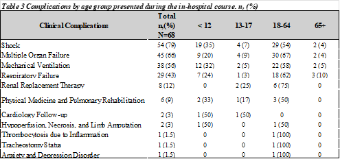
The most frequent complications after the 4-day onset of symptoms were shock, multiple organ failure, and the need for mechanical ventilation, respectively, affecting the 18-64 year age group and children under 12 years of age. Table 3.
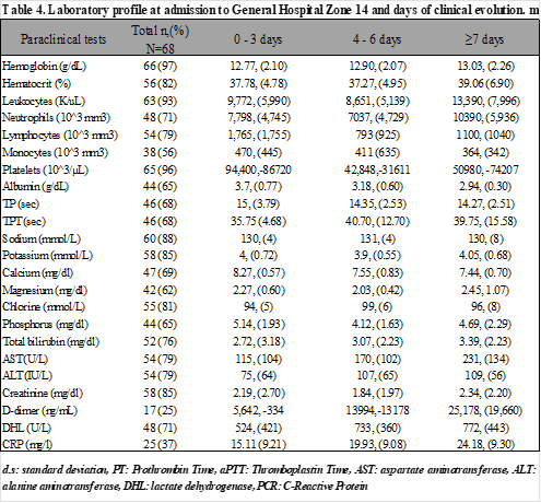
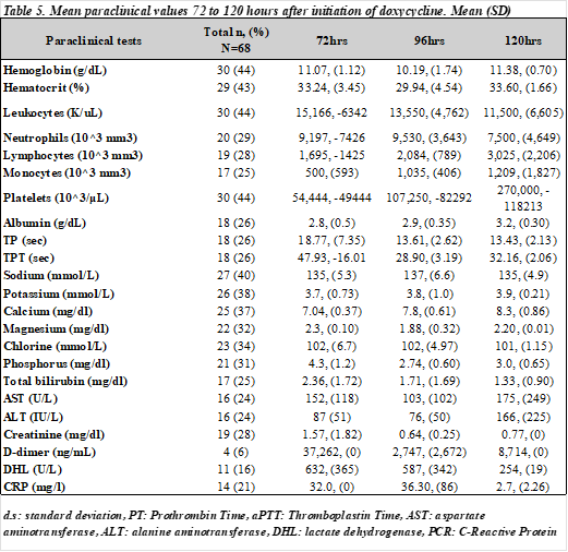
The mean and standard deviation of laboratory results available upon patient admission were calculated according to the number of days since symptom onset. Abnormalities were observed from the first 3 days after symptom onset, including neutrophilia, thrombocytopenia, hypoalbuminemia, hyponatremia, hyperbilirubinemia, and elevated transaminases, creatinine, D-dimer, lactate dehydrogenase, and C-reactive protein. Table 4. Hematological follow-up was performed between 72 and 120 hours after the initiation of doxycycline in patients hospitalized for more than 5 days with available paraclinical results. In general, a trend toward normalization was observed compared to the abnormalities noted upon admission. It should be noted that two of these cases resulted in a fatal outcome. Table 5.
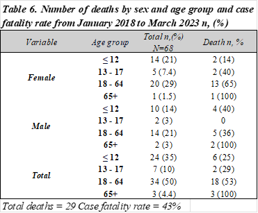
Finally, the case fatality rate during the study period was 43%, with the female population between 18 and 64 years of age being the most affected, representing 65% (including 1 patient with a pregnancy of 11.5 weeks of gestation), followed by the pediatric population < 12 years of age with 6 deaths, corresponding to a case fatality rate of 25% in this age group (Table 6).
DISCUSSION
A descriptive study was conducted on a total of 68 confirmed cases of Rocky Mountain Spotted Fever. The results show a considerable percentage of cases with an epidemiological history of tick exposure, based on the presence of ticks in and around the patients' homes and tick bites within 14 days prior to symptom onset. However, in 18% of cases, the history of exposure was unknown. This finding is important to consider, as it aligns with the literature, which indicates a high percentage of cases lacking this history.8
Furthermore, the analysis in this study observed symptom onset between 2 and 8 days after the tick bite, which differs from the literature's description of symptom onset as 4 to 10 days post-bite, even though it coincides with the incubation period. Clinical manifestations vary according to the different stages of the patient's illness. During the first three days of illness, signs and symptoms such as fever, headache, and rash, accompanied by gastrointestinal symptoms, predominated. However, when analyzing the classic triad as a set of signs and symptoms (fever, rash, and headache), only 38% of patients presented with this triad, a lower percentage than described in the literature.8 It is important to recognize that this study yielded nonspecific symptoms of the disease, as described in the literature, and these may represent other differential diagnoses. Likewise, during the course of the illness, other signs and symptoms may appear and not be present at the time of diagnosis, thus missing the opportunity for timely treatment. That is, in the first 72 hours of the onset of clinical symptoms, a considerable percentage of patients developed gastrointestinal symptoms, which coincides with the frequency of medical consultations prior to admission.
Differential diagnoses such as gastroenteritis and pharyngitis, especially in the pediatric population, predominated, and inadequate treatment was administered.
After four days, symptoms progressed to dyspnea, neurological impairment, signs of third-space fluid leakage, active bleeding, and hepatic and renal failure—processes related to the complications described in the literature.9 Among the initial laboratory tests, thrombocytopenia, hyponatremia, and elevated liver enzymes were findings that contributed to the suspicion of the disease, and these were analyzed and highlighted in this study. Seventy-nine percent of the patients presented with shock, along with other complications that arose after admission (four days after the onset of symptoms), such as multiple organ failure, the need for mechanical ventilation, respiratory failure, and renal replacement therapy. Consequently, some patients presented with sequelae at discharge, including pulmonary rehabilitation, hypoperfusion, necrosis, and limb amputation, as reported in the literature.10 Doxycycline was most frequently administered after the first six days of illness, which may have been a determining factor in the patients' fatal outcomes, considering studies conducted in Mexico that have shown that administering treatment after 72 hours from the onset of fever results in a sevenfold increase in the probability of a fatal outcome.11
Once the specific antibiotic was administered, laboratory profiles were monitored between 72 and 120 hours in cases that remained hospitalized for more than five days to assess changes in parameters. All laboratory results were collected from the medical records of all cases upon admission and for follow-up. 27 cases had a fatal outcome upon admission and at 24-48 hours, 3 patients spent more than 72 hours without hematological follow-up, 4 cases were outpatients and 4 were transferred to another unit so it was not possible to include this group in the hematological follow-up results.
In the remaining patients, the same paraclinical indicators were found to be affected, improving during their clinical course. These improvements may have been observed in those patients who recovered without sequelae, while those who remained abnormal were patients with a fatal outcome or who recovered with sequelae. The case fatality rate remains high in the state of Sonora, reaching 50% in 2022 alone and 43% in the population admitted to this hospital between 2018 and 2023. Limitations included the diagnosis based on only one positive serological test, although these patients presented with clinical symptoms suggestive of Rickettsiosis. However, in accordance with the National Guidelines for Epidemiological Surveillance and Rickettsiosis Laboratory testing, these patients were excluded from this study. Another limitation is the incomplete information available for monitoring the laboratory profiles of patients who began treatment upon admission, for the reasons explained.
The findings of this study contribute to providing information on the epidemiological background of patients, since, as reported in the literature, there are cases where exposure to the vector is unknown. Given that Sonora is an endemic state for FMMR, this finding should not preclude a suspected diagnosis, and these patients should be closely monitored. It is important to enrich the existing knowledge of healthcare personnel regarding FMMR and to inform the community about its symptoms, which in this research are nonspecific due to the characteristics of its evolution and presentation. Furthermore, this study provides information on the stage of the disease in patients, along with the laboratory parameters most frequently affected upon admission, to offer better guidance when a diagnosis is suspected and to provide timely treatment.
In this study, 68 cases were considered, all classified as confirmed by tests recognized by the InDRE (National Institute of Epidemiological Diagnosis and Reference) as outlined in the 2022 National Guidelines for Laboratory Surveillance of Rickettsiosis. The null hypothesis was accepted, given the lack of specific epidemiological and clinical characteristics in most patients diagnosed with FMMR (Fascicle-Mighting-Related Rickettsial Diseases), as the prevalence of the classic triad was less than 50%, and accompanying symptoms predominated. However, the reported complications and laboratory parameters showed alterations consistent with those described in the literature. A case fatality rate of 43% was observed, with adults being the most affected, followed by children under 12 years of age, who accounted for 25% of deaths.
REFERENCES
1.Álvarez-Hernández G, Roldán JFG, Milan NSH, Lash RR, Behravesh CB, Paddock CD. Rocky Mountain spotted fever in Mexico: past, present, and future. Lancet Infect Dis. 2017 Jun;17(6):e189–96
2.Stewart AG, Stewart AGA. An Update on the Laboratory Diagnosis of Rickettsia spp.Infection. Pathogens. 2021 Oct 13;10(10):1319.
3.Murray PR, Rosenthal KS, Phaller MA. Capítulo 34 Rickettsia, Ehrlichia y bacterias relacionadas. Microbiología Médica. 2017. 8ed. 338-347.
4.Reyes-Castro PA, Ernst KC, Walker KR, Hayden MH, Alvarez-Hernandez G. Knowledge, Attitudes, and Practices Related to Rocky Mountain Spotted Fever in Hermosillo, México. Am J Trop Med Hyg. 2021 Jan 6;104(1):184–9.
5.Razzaq S, Schutze GE. Rocky Mountain Spotted Fever. Pediatr Rev. 2005 Apr 1;26(4):125–30.
6.Walker DH, Ismail N. Emerging and re-emerging rickettsioses: endothelial cell infection and early disease events. Nat Rev Microbiol. 2008 May;6(5):375–86.
7.Martinez EB, Mendoza AP, Marquecho FM, Plata MC, Verdugo MR. Letalidad por fiebre manchada por Rickettsia rickettsi en pacientes de un hospital pediátrico del estado de Sonora, 2004-2012. 2013 Marzo;55(2):151-152.
8.Braun DS, Greenberg I, Pagadala M. Rocky Mountain Spotted Fever Masquerading as Gastroenteritis: A Common but Overlooked Clinical Presentation. Cureus [Internet]. 2021 Apr 12 [cited 2022 Dec 5]; Available from: https://www.cureus.com/articles/ 53812 -rocky-mountain-spotted-fever-masquerading-as-gastroenteritis-a-common-but-overlooked-clinical-presentation.
9.Álvarez-López DI, Ochoa-Mora E, Nichols Heitman K, Binder AM, Álvarez-Hernández G, Armstrong PA. Epidemiology and clinical features of Rocky Mountain
10.Snoweden J, Simonsen KA. Rickettsia Rickettsiae. StatPearls Publishing; 2022
11.Rodríguez-Muñoz L, Barrera-Salinas R, Sánchez-García C, Solórzano-Santos F, Vaquera-Aparicio DN, López-Castillo D. Rickettsiosis de fiebre manchada. Estudio de casos notificados en un hospital pediátrico de segundo nivel en el noreste de México, 2012-2022. Gac Med Mex [Internet]. 2023;159(2). Available from: http://dx.doi.org/10.24875/gmm.22000354
Citation
Cantú-González SP and Serrano-García EA, Epidemiological characteristics in patients diagnosed with Rocky Mountain Spotted Fever, ERSJ 2026,2(1) 28-40
Academic Editor: Porfirio Felipe Hernández-Bautista, Received: April-27-2026, Revised: June-30-2026, Accepted: July-06-2026, Published: July-18-2026.